Anterolateral Thigh Free Flap for Gustilo IIIB Open Tibial Fracture with Segmental Soft-Tissue Loss
Delayed reconstruction of a 18 × 12 cm anterolateral leg defect with exposed tibia and hardware after high-energy trauma, using a perforator-based ALT free flap and end-to-side microvascular anastomosis to posterior tibial vessels.
Patient Information
- Age: 34 years
- Gender: Male
- Diagnosis: Gustilo IIIB open tibial fracture with segmental soft-tissue loss, exposed plate, and infected wound bed after external fixation
Procedure
Staged orthoplastic reconstruction: radical wound débridement, targeted muscle and bone culture—guided antibiotics, vacuum-assisted closure (VAC), and definitive anterolateral thigh (ALT) free fasciocutaneous flap coverage with simultaneous internal fixation revision where indicated.
Intraoperative Images
Findings
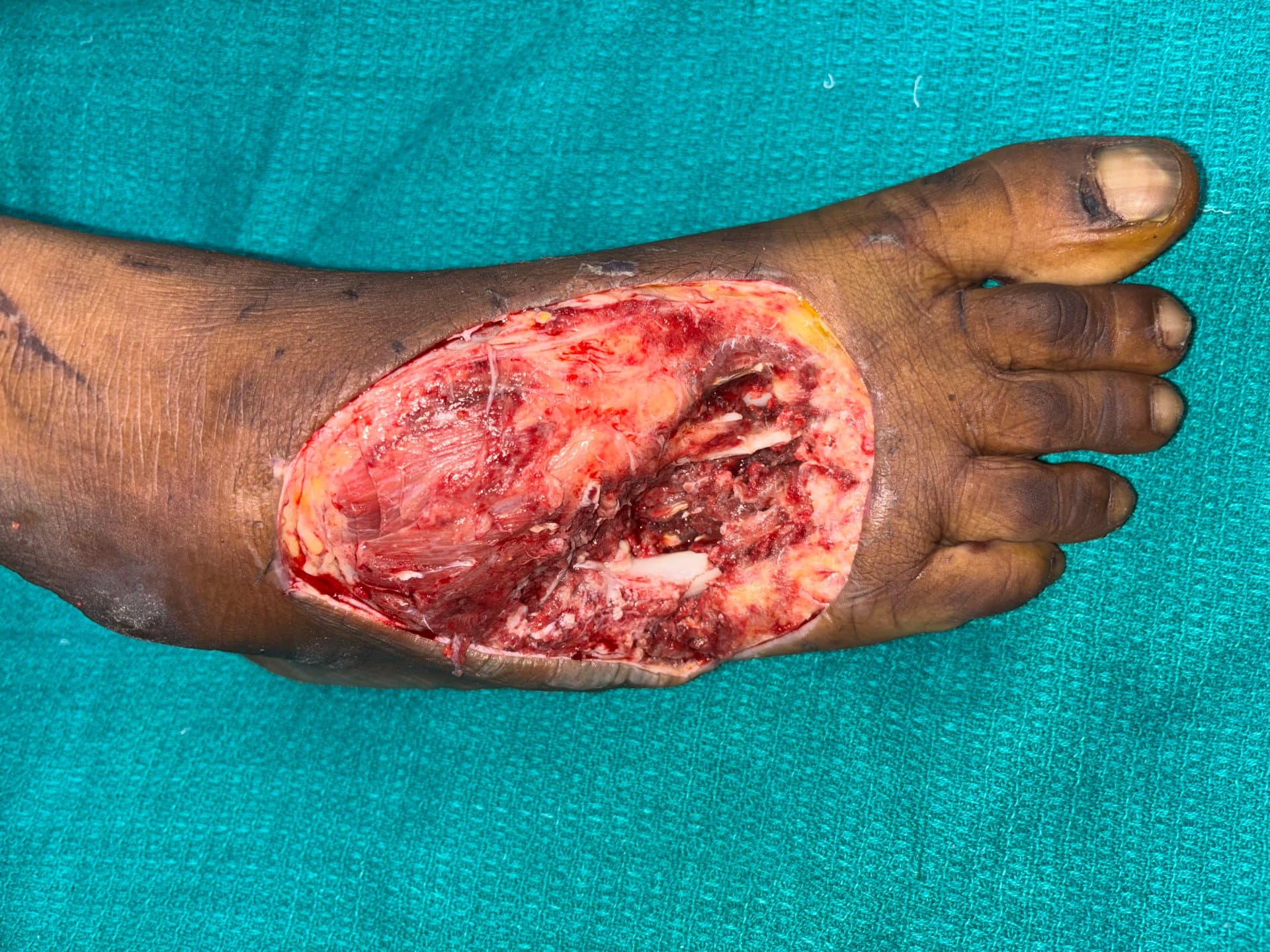
On presentation for definitive reconstruction, the patient had a chronic 18 × 12 cm defect over the anterolateral distal third of the leg with exposed tibial cortex and orthopaedic hardware, indurated periwound skin, and bridging granulation only at the proximal margin. Angiographic and handheld Doppler assessment confirmed patent posterior tibial and peroneal recipient vessels; the anterior tibial artery was compromised from initial injury. Preoperative workup included normalized inflammatory markers, negative deep tissue culture after débridement, and haemoglobin optimisation above 10 g/dL.
Surgical Technique
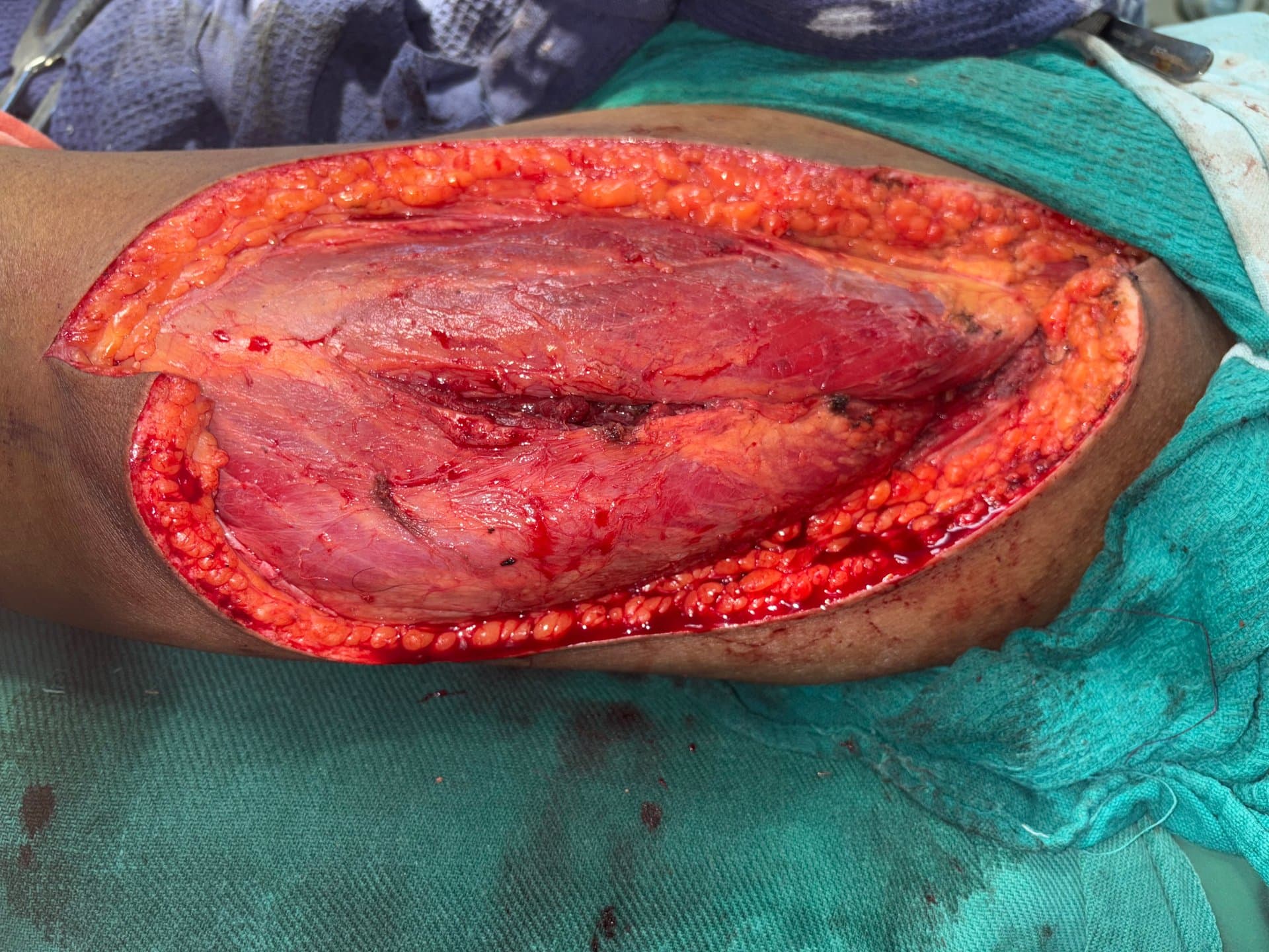
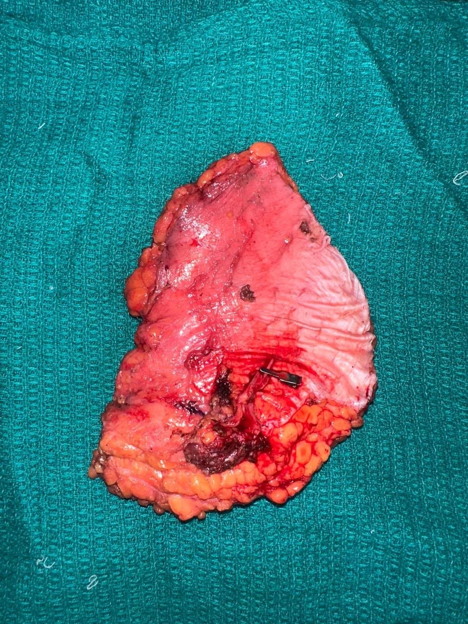
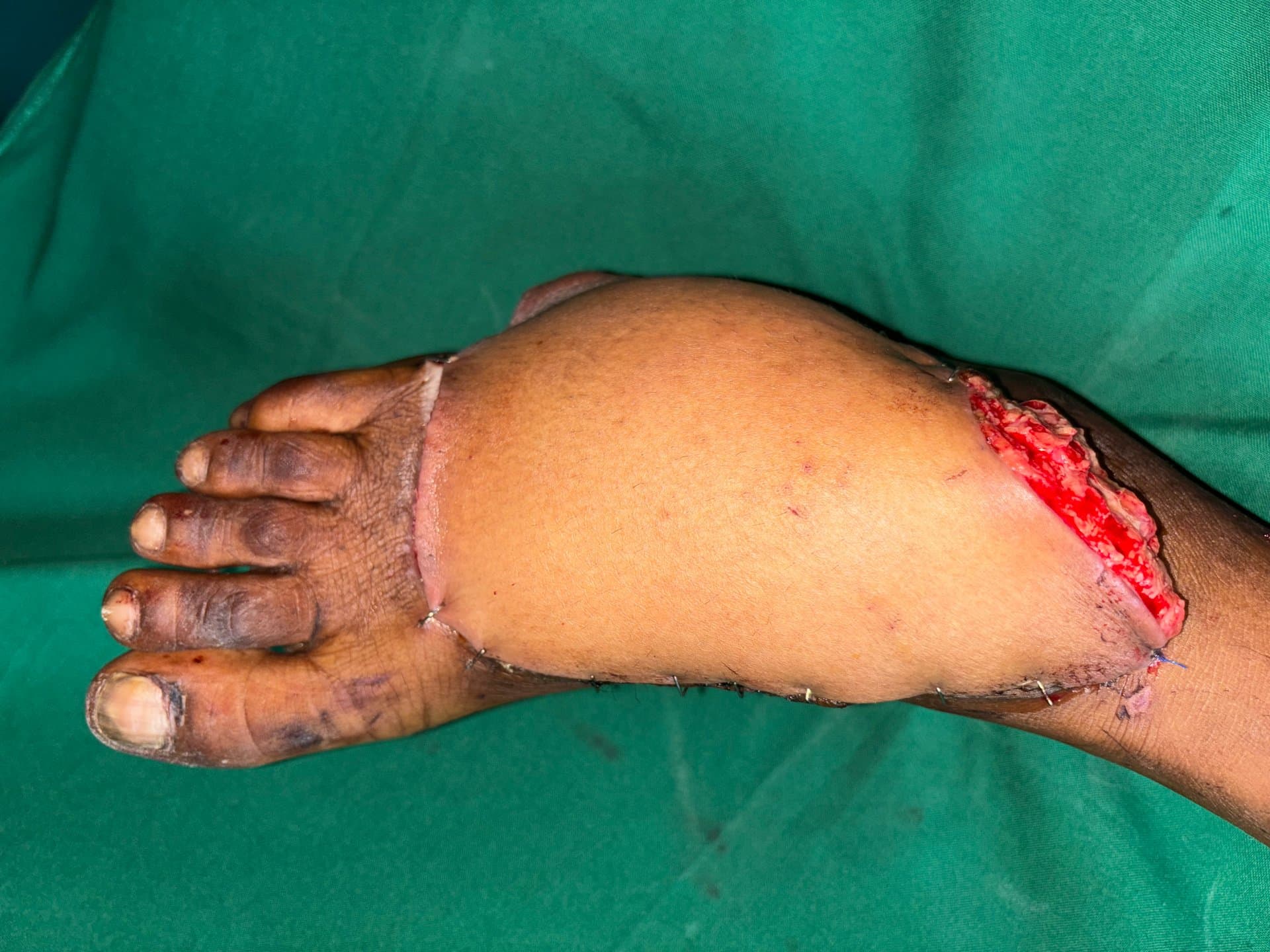
Under general anaesthesia with tourniquet control where appropriate, recipient bed preparation included excision of scarred, non-adherent tissue, cortical drilling of exposed bone to encourage bleeding, and removal of biofilm-colonised hardware segments as per orthopaedic plan. A left ALT flap was designed along a line from the anterior superior iliac spine to the lateral patella, centred on a single dominant perforator from the descending branch of the lateral circumflex femoral artery identified with handheld Doppler. Flap dimensions were 20 × 13 cm including a 3 cm adipofascial cuff around the perforator. The pedicle was dissected to adequate length with two venae comitantes and the primary perforator preserved. Recipient vessel preparation on the posterior tibial system included adventitial stripping and topical papaverine. Arterial anastomosis was performed end-to-end with 9-0 nylon under operating microscope; venous anastomosis used a 2.0 mm coupler on the dominant vena comitans. Flap inset was tension-free with suction drains and a bolster dressing over the skin paddle. Postoperative protocol included ICU monitoring for 48 hours, hourly flap checks (colour, capillary refill, temperature, Doppler signal), low-molecular-weight heparin per unit protocol, and strict limb elevation.
Outcome
Flap remained viable with brisk capillary refill and audible arterial Doppler signal throughout the inpatient stay. Partial distal margin superficial epidermolysis at day 9 managed conservatively. At 6-week follow-up, complete flap survival, stable soft-tissue envelope over hardware, and progression to partial weight-bearing per orthopaedic guidance. At 3 months, the patient ambulated with support, donor site healed with acceptable contour, and no osteomyelitis recurrence.
Clinical Notes
Highlights the importance of staged orthoplastic management—infection control and recipient vessel assessment before free tissue transfer. Add hero and intraoperative images to public/journals/ (e.g. alt-free-flap-hero.jpg, alt-free-flap-inset.jpg) when available.